Establishing Primary Neuronal Cultures
from Drosophila Pupae
Designing a neurobiological assay for synaptic development
Overview
Goal
Develop primary neuronal cultures from Drosophila melanogaster pupal brains to create a controlled assay for studying synapse formation.
Approach
Late stage pupae (55 to 78 hours) underwent brain dissection, papain based enzymatic dissociation, and culture in DDM2 medium following an adapted Sicaeros et al. (2007) protocol.
Outcome
Cultures did not produce viable neurons due to contamination and low cell yield, but the experiment revealed critical methodological insights for future iterations.
The Problem
Studying synaptic development in vivo limits control over experimental variables. In vitro neuronal cultures offer manipulation and homogeneity, but bridging the gap between a living brain and a culture dish introduces significant procedural challenges.
From Brain to Culture
In Vivo Brain
Intact pupal CNS
Dissection
Brain isolation
Dissociation
Enzymatic digest
Culture
DDM2 medium
Analysis
Microscopy
Genetic Tractability
GAL4/UAS system enables precise genetic screening and manipulation of neural circuits.
Low Cost, High Scale
Short life cycle, small genome, and ease of maintenance make Drosophila ideal for high throughput assays.
Disease Modeling
Drosophila models successfully replicate aspects of human neurological disorders for pharmacological screening.
Approach
Specimen Selection
10 to 15 golden brown Drosophila pupae aged 55 to 78 hours post pupariation. At this stage, pupae are developmentally ready for optic lobe dissection and CNS extraction.
Dissection
Cuticle removed with 27 gauge syringe needle. Head extracted by severing the neck with forceps. Brain isolated by creating a vertical incision through the forehead and pushing the brain out with a needle.
Enzymatic Dissociation
Brains transferred via 20 uL pipette into papain solution (0.8 mg 100X cysteine, 35.7 uL papain, 150 uL DS cysteine, 5 mL DS buffer). Incubated on rotator at 60 rpm for 10 minutes, then centrifuged at 1600 rpm for 3 minutes.
Culture Setup
Resulting material placed in DDM2 culture medium (DMEM, insulin transferrin selenium, hydrocortisone, progesterone, putrescine). Incubated in CO2 incubator.
Observation
Cultures examined via dissecting microscope at 4X magnification after 4 to 8 days of incubation. Assessed for neuronal growth, contamination, and cell viability.
Experimental Iteration
Two trials tested and refined the protocol. Each failure produced specific diagnostic information that shaped the next attempt.
Trial 1
Conditions
Non sterile dissection (no laminar flow hood). Standard papain digestion timing. 7 to 9 brains collected.
Result
Fungal contamination after 4 days. Green fibrous growths and medium discoloration. Cultures discarded.
Trial 2
Adjustments
Improved sterility protocols. 6 brains dissected (2 sets of 3). Split digestion timing: 10 min vs. 5 min. Two DDM2 washes added.
Result
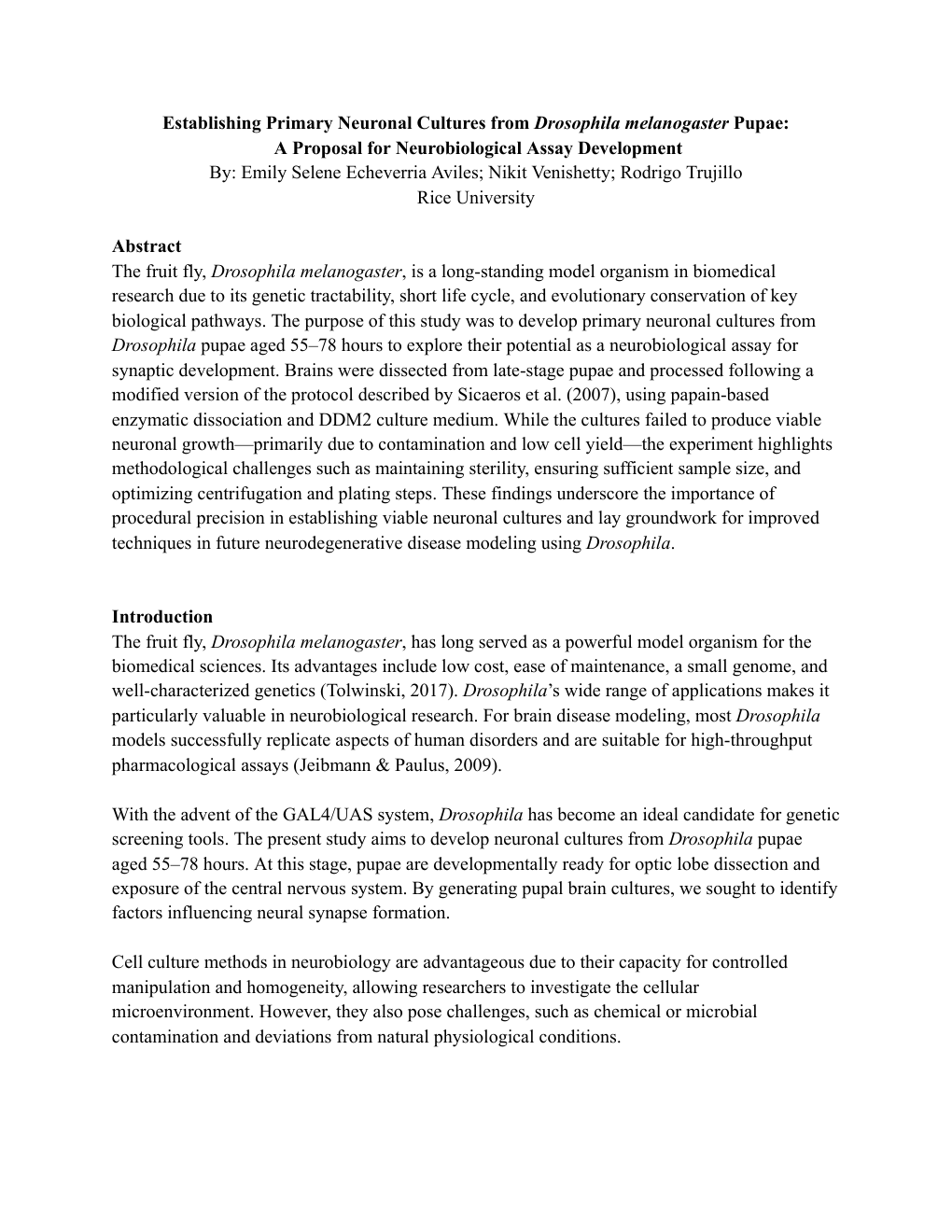
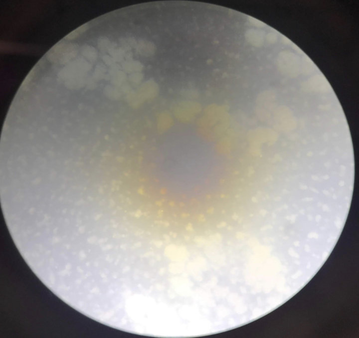
Contamination persisted. Central green mound with brown discoloration. White cellular aggregates visible but likely nonviable debris.
Results
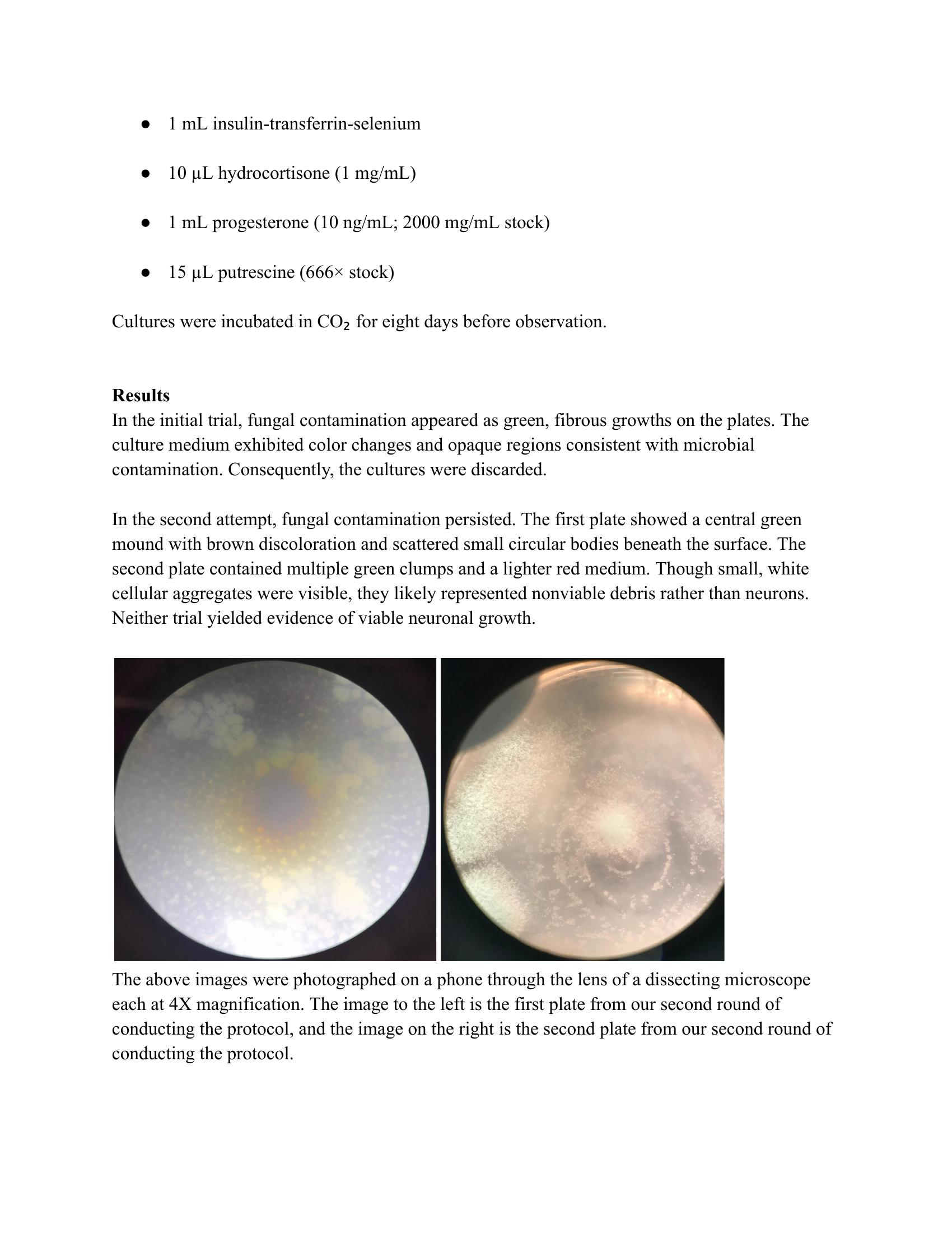
Neither trial yielded evidence of viable neuronal growth. The microscopy images below show the contaminated culture plates from the second trial at 4X magnification.
Trial 2 Culture Plates
Left: Plate 1 showing central green fungal mound with brown discoloration and scattered circular bodies beneath the surface. Right: Plate 2 with multiple green clumps and lighter red medium indicating contamination.
Green Fungal Growth
Fibrous green growths appeared on both plates, indicating microbial contamination introduced during dissection or plating.
Medium Discoloration
Color changes in culture medium from clear pink to opaque yellow/brown, consistent with microbial metabolic activity.
No Viable Neurons
Small white cellular aggregates appeared but likely represented nonviable debris rather than neuronal growth. No neurite extension observed.
Key Insights
The failure to culture viable neurons illuminated three specific procedural factors that must be addressed for future success.
Sample Size Matters
The reference protocol calls for 10 to 15 brains per sample. Only 7 to 9 brains collected in Trial 1, reducing neuronal yield below the viability threshold. Separating optic lobes from central mushroom bodies may have further compounded neuron loss.
Cell Recovery Failure
No visible pellet formed after centrifugation at 1600 rpm for 3 minutes, indicating insufficient cell concentration for proper washing and plating. Without a concentrated cell mass, downstream steps were compromised.
Sterility is Critical
Conducting dissections outside a laminar flow hood introduced fungal contamination that persisted even after procedural adjustments. All dissection and culture steps must occur under strict sterile conditions.
What Would Change in Future Iterations
Increase sample size to full 10 to 15 brains per trial
Optimize centrifugation parameters until a visible pellet forms
Enforce strict laminar flow hood sterility for all steps
Standardize enzymatic digestion timing with controlled experiments
Impact and Learning
Experimental Design
The project demonstrated systematic experimental design: hypothesis formation, protocol adaptation, controlled variable testing, and diagnostic analysis of failure modes.
Failure Diagnosis
Each trial produced specific, actionable findings rather than ambiguous results. The ability to diagnose failure precisely is as valuable as achieving success.
Iterative Refinement
Trial 2 incorporated targeted adjustments based on Trial 1 observations: improved sterility, split digestion timing, and additional wash steps.
Foundation for Future Work
The findings lay groundwork for improved Drosophila neuronal culture techniques applicable to synaptogenesis, neural circuitry, and neurodegenerative disease modeling.
Research Significance
Methodological refinement and procedural precision determine whether in vitro systems can reliably model in vivo biology. Failure, diagnosed precisely, accelerates progress more than ambiguous success.